Friday, July 20, 2012

Lesson eight: DNA packaging


Lesson eight: DNA packaging

In the previous lessons we had discussed Transcription and Translation.  As previously stated, the process of Transcription converts a DNA sequence into an mRNA.  The mRNA is then converted into a protein through the process of Translation.  The complexity of Transcription and Translation is so immense that decades and millions of dollars dedicated towards researching these two concepts was and still is performed today.  These two principles make up the backbone of molecular biology, but an additional secondary important concept is DNA packaging.

There is about 3 meters of DNA in human cells.  The entire DNA molecule has to fit into cells of about 10 to 100μm (the “μ” symbol means micro- which is 10-6).  This is like taking a string from Bloomington to the outskirts of Columbus, IN which is 30 miles away.  Exactly how this entire amount of DNA is packaged into cells is still being researched, but scientists have discovered some basic levels of packaging.

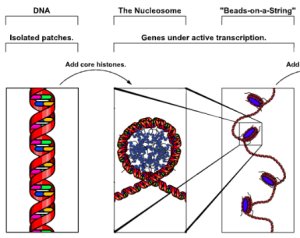
The first level of packaging is called the 10nm “Beads on a string” (10nm means 10 nanometers and the prefix nano- means 10-9).  This level is exactly how it sounds: it is 10nm wide and looks like “Beads on a string”. In this level DNA is wrapped around a complex but important protein complex called a histone.  The histone comprises the “beads” portion and the DNA the string portion of the “Beads on a String” term.  The backbone of DNA is negatively charged and the histones are made up of positively charged amino acids, thus this serves as the basis of how the histones interact with the DNA.  When DNA is wrapped around a histone is called a nucleosome.

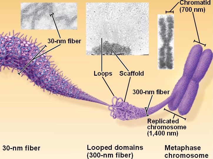
The next level of packaging is called the 30nm fiber.  In this level the 10nm “Beads on a string” is folded upon itself many times over to form a structure that is 30nm thick.  In this level there are six nucleosomes per turn that look like a bunch of beads packaged into a cylindrical shape.

The final known level of DNA packaging is only visible when cells are dividing.  At a particular stage during cellular replication the DNA becomes so condensed that it is visible under a microscope.  This particular structure is called chromatin.  When cells are not actively dividing the DNA is not fully condensed and not directly visible under the microscope.  The exact mechanism of this packaging is still being studied.  In fact, all mechanisms of DNA packaging are still being currently studied.


Here are some Youtube videos discussing the concept of DNA packaging:


Monday, July 16, 2012

Seventh Lesson: Overview of Proteins


Seventh Lesson: Overview of Protein Folding

From the previous lesson we have learned that through the combination of both Transcription and Translation, DNA is converted into proteins.  Remember that proteins are made up of amino acid building blocks that are attached through peptide bonds (a special type of covalent bond). Proteins become functional as the amino acids naturally fold into the right native structure as they emerge from the ribosome.  Only when a protein is folded correctly is it functional.  Why?  Think about it this way.  Many proteins work through a lock & key mechanism in which something binds and the protein converts it into something else.  A lock has to be the right shape to fit its particular key.  If a lock is not the right shape (a protein that is improperly folded), it will not work correctly.

There are four levels of protein complexity that range from simple to the most complex: primary, secondary, tertiary, and quaternary.  All proteins have some form of a primary, secondary, and tertiary folding, but not every protein has quaternary structure.

The primary structure is simple.  If I were to read to you each amino acid in a series of connected amino acids to you in order, you would have the primary structure.  It is like reading the sentences in a book in order.  See, I told you it was simple, right?

The secondary structure is a little bit more complex than the primary structure.  In the secondary structure the linear backbone of the protein begins interacting and taking shape.  Amino acids will fold to form two different but common structures: either α-helices or β-strandsα-helices are helical structures formed by the amino acid backbone interacting together (something that looks like a screw).  β-strands are structures that occur when the backbone folds on top of itself and interacts with hydrogen bonds.  (The picture below helps a lot to visualize it.)


The tertiary structure is the last universal protein structure.  The tertiary structure, by definition, is the 3D arrangement of all the amino acids of the proteins.  In other words, it is the 3D image of the entire protein.  This is the most complex and most common image we see of most proteins today.  Most tertiary structure images are found on very powerful computers that are designed to predict protein folding.
The term protein is very misleading; many proteins have multiple but distinct chains of amino acids that fold together to form a bigger structure.  Think of it this way: it is like teeing a bow. A bow can be constructed from one piece of string, but more complex bows use multiple pieces of string to form the structure.  When this happens, the protein is said to have a quaternary structure.  Not ever protein is comprised of many different individual chains of amino acids, so some natural proteins only have a tertiary structure. (Many molecular biologist use the term polypeptide to identify a protein that is comprised of only a single chain of amino acids and not more than one.)  In the next lesson I will be discussing the concept of DNA packaging.  This involves taking an object that is 3 meters long and stuffing it into a space of that is only fractions of a hair thick!

Here is are some Youtube videos discussing protein folding


Sunday, July 15, 2012

Sixth Lesson: Translation continued


Lesson 6: Translation continued.

In the previous lesson we began to discuss the concept of Translation.  Remember, Transcription involves taking the nucleotide gene sequence found in the DNA and converting it into an mRNA molecule.  Translation is performed next and changes the mRNA that was originally defined by the DNA into a particular protein sequence.  Through the combination of these two processes, DNA is converted into proteins.  The main mechanistic workhouse of Translation that performs the vast majority of the work is called the ribosome.

Before going into more detail about Translation, I need to discuss some basic protein chemistry.  The topic of the next lesson is going to cover proteins in much more detail, but for now let us discuss some simple pointers.  Like DNA, which is made up of small building blocks called nucleotides, proteins are also made up of small building blocks called amino acids.  In either case, these building blocks are like Lincoln Logs, in which they are simple pieces that come together to build a larger structure.  There are 20 different types of amino acids found in nature, and amino acids bond together with a special type of covalent bond called a peptide bond. (See Lesson 2: Chemical Bonding if you are confused about the term covalent bonding. )

Translation occurs in the ribosome (just like the building of a car occurs in a factory) but requires another special class of RNA molecule called a tRNA.  tRNA’s serve as important players during Translation.  (Remember that RNA and DNA are both made up of nucleotides.) Together the ribosome and tRNA’s function to take the mRNA molecule and convert it into a protein sequence.

Basically, take the mRNA molecule and count every three nucleotides, (1, 2, 3…1, 2, 3….1, 2, 3…).  For every triplet you counted, this is called a codon.  Remember tRNA’s?  Well, there are many different types of tRNA molecules and for each molecule there is a very important region found on them comprised of three nucleotides also.  This is called the anticodon.  The anticodon of a given tRNA matches a particular codon of an mRNA through complementary base pairing rules.  For example, if a given codon on the mRNA is GAU then the complementary anticodon of the tRNA is CUA.

G  ->  C
A  ->  U
U  ->  A


Every possible combination of three nucleotides (codon) on a mRNA has a matching anticodon sequence on a tRNA.  In other words, there is a given tRNA molecule that contains a matching anticodon for every possible codon combination (there are 64 mathematical combinations).

The importance of the anticodon-codon base-pairing combination is due to the fact that this is the means the cell uses to distinguish what is supposed to be the correct amino acid building blocks to use.  Amino acids are attached to tRNA’s.  A particular amino acid will be found on only the right tRNA with the right corresponding anticodon. 


Yet, why are there 64 codon-anticodon combinations but only 20 amino acids?  The simple answer to this question is that there are more than one codon-anticodon combinations that encode for the same amino acid.  For example, the amino acid glycine is encoded by either 4 different codons: GGU, GGC, GGA, and GGG.  The term molecular biologist use is called "redundant" or "redundancy" to explain this concept.  You can use this table to visual this for yourself:


There are not that many great analogies I can use to teach this concept that is why I am providing a bunch of YouTube videos for reference.

Reference YouTube videos:

A fun cartoon animation featuring Mario Brothers!
http://www.youtube.com/watch?v=ZPlnDzkBrpc&feature=endscreen&NR=1

A good visual but a little complex:
http://www.youtube.com/watch?v=TfYf_rPWUdY&NR=1

Also a good visual a bit on complexity: 
www.youtube.com/watch?v=1PSwhTGFMxs&NR=1&feature=endscreen

Wednesday, July 11, 2012

Fifth Lesson: Transcription Review and Translation Introduction


As we remember the “mechanistic work” of the cell is performed by proteins, yet the information to build proteins is encoded in the nucleotide sequence of DNA.  How do we take the data present within the nucleotide sequences of DNA into proteins?  Previously, we had discussed that there is an intermediate involved in this process called mRNA.  Only one of the strands of DNA contains the information required to build a functional protein (because of course, the DNA strands are complementary due to base-pairing rules).  A single-stranded molecule that is complementary to the protein-defining strand is first formed and this is the mRNA.  In other words the mRNA is complementary to the particular one strand in the DNA that contains the information required to form the given protein.  (In reality molecular biologists know exactly which strand of DNA is involved in protein formation and which strand is exactly considered the complementary strand that matches the exact sequence of mRNA.)  The process of forming this complementary strand is called Transcription.  (See Lesson 4 for more information)


After the mRNA molecule is formed from Transcription, the next processed involved is called Translation.  Translation involves taking the sequence of the mRNA molecule and converting it into a protein.   Translation is performed by a very complex molecule comprised of both a special, different type of RNA, called rRNA, and proteins.   Together both the rRNA pieces and protein pieces form what is known as the ribosome.  Think of the ribosome has a hybrid engine: it has protein parts and rRNA parts that work together to form a functional unit, just like a hybrid engine usually has gasoline and electric parts that work together to produce energy.  The ribosome takes the information in the mRNA and converts it into a new protein.  This is a very complex process that I am going to divide between two sections, thus I will continue discussing this topic in the next section.


(The color portions are the protein part and the grey part are the rRNA part)


Additional Videos for Reference:
(Some of these maybe a little bit complicated but still offer visualizes for the points I make)

http://www.youtube.com/watch?v=Jqx4Y0OjWW4&NR=1

or


Sunday, July 8, 2012

Fourth Lesson: Transcription


In the last lesson we discussed the importance of DNA and the concept of a gene.  Genes encode for all the functional "stuff" of the cell but do not directly participate in the work of cells' life-cycles.  Think of it this way.  If a cell can be thought of as a car, the car is made up of various different parts that all need to work in unison and at the right time, like the engine, brakes, ignition, etc.  Likewise, a car is defined by a set of instructions that tell you have to make the car; in this case it is the car's blueprint.  The blueprint does not directly tell the car how to function but instead indirectly serves as a guide for all the other components to tell them how to work and come together.  This is the case for DNA.  DNA does not do any of the “mechanical” work of the cell, but most commonly indirectly encodes for proteins, which in turn serve as the “nuts and bolts” of the cell.

The conversion of a DNA gene sequence into a protein occurs through an intermediate.  This intermediate molecule is called messenger ribonucleic acid or mRNA.  mRNA like DNA is considered a nucleic acid and is made up of nucleotides, though the nucleotides for DNA and RNA are slightly chemically different.  For instance, instead of thymine in DNA, mRNA contains uracil in all places where thymine would have been found in the DNA the mRNA originated from.  There are many types of RNA (and all are single-stranded unlike double-stranded DNA), but I am only going to talk about mRNA.


As I have stated before, the genetic information of DNA lies in the sequence of the four nucleotides: adenine, thymine, cytosine, and guanosine.  (I have chosen to provide a bit of a visual for this lesson below.) This genetic information in the DNA is converted into mRNA in the process called Transcription.  mRNA serves as a complementary template according to the nucleotide base-pairing rules of the gene on the DNA that it is encoding from.  For example if the gene sequence is ATCG, then the complementary mRNA sequence would be TAGC.  The entire gene is converted into its complementary sequence into the mRNA intermediate. The mRNA is then used to tell the cell how to make a given protein in the process of Translation, which I will be discussing next.

Base Pairing Review:

A ->  T
T  -> A
G ->  C
C ->  G

*The letters on the first column represent the four nucleotides that are found in DNA.  The letters in the second column with the arrow pointing towards them indicate the nucleotides that will base-pair with the frist column.  You can also see the Third Lesson for more review.

The Gene Example:

A --  T
T --  A
C --  G
G --  C

Notice that the gene in the DNA is double stranded!

The mRNA complement from the gene would be:


A --  T                                              - U
T --  A                  -------->                 - A
C --  G                                              - G
--  C                                              - C

Notice that the mRNA is single-stranded and complementary to the gene sequence, and therefore matches up perfectly with one of the DNA strands!  Also notice that thymine is replaced by uracil!

Reference Videos:
Some Videos covering the Topic of Transcription:


Monday, July 2, 2012


Third Lesson: DNA overview


Now that we have discussed chemical bonding, we have a solid footing to begin discussing the fundamentals of molecular biology.  In this lesson I am going to discuss DNA.  DNA serves as probably the most important concept in molecular biology, because it defines all important biological processes.


DNA stands for Deoxyribonucleic acid.  Francis Crick and James Watson were credited with the discovery of DNA, and received the Nobel Prize in Medicine or Physiology in 1962.  DNA is described as two long strands wrapped around each other to form a long double-helix 

The building blocks of DNA are called nucleotides.  There are two categories of nucleotides: pyrimidines and purines.  Furthermore, there are two types of pyrimidines used in DNA, and these are cytosine and thymine.  There are two types of purines used in DNA, and these are adenine and guanine.


A given adenine nucleotide found on one strand of DNA will form two hydrogen bonds with a thymine nucleotide on the opposite strand and vice versa.  Likewise, a given guanine molecule found on one strand of DNA will form three hydrogen bonds with a cytosine molecules on the opposite strand and vice versa.  The unique way all the nucleotides pair together leads to the formation of the double-helix.


The importance of DNA lies in the unique sequence that the nucleotides are arranged in on the strands of DNA.  Every living thing on the planet’s distinctive linear sequence of nucleotides makes that organism what it is.  Humans, bacteria, plants, etc. all are defined by DNA sequences.  Within DNA there are important areas that encode and define how to make all the biological material in the world.  These areas are called genes.  There are two processes involved in which gene sequences on DNA are converted into the functional biological products that do the “work” of molecular biology.  First Transcription followed by Translation, which I will be discussing in the next upcoming lessons.

Wednesday, June 20, 2012

Second Lesson: Chemical Bonding


Greetings to all you curious science minds!  Today I would like to continue off of the previous post's topic and discuss some more basic chemistry.  Again, I am always surprised by how little this is understood between biology students.  (This is made especially evident nearly every time I have tutored for organic chemistry).


Since we now have a basic understanding of what exactly an atom is, we can now discuss how they combine to form molecules that make up the basis of everything around us.  If an atom loses an electron, which of course is negatively (-) charged, then the atom is said to be positively charged (+).  Likewise, when a given atom loses an electron that electron is gained by another atom which makes that atom overall negatively charged (-).  Protons and neutrons are typically not transferred as easily as the small readily displaced orbiting electrons are.  


Any atom that has a charge is called an ion.  When two ions of opposite charge meet they are attracted to each other and will weakly bond together.  This type of bond is called an ionic bond

The more common type of bond used in biology is a covalent bond.  The reason why a covalent bond is more common is because a covalent bond is significantly stronger than an ionic bond.  Basically in a covalent bond, electrons are shared between two atoms instead of being transferred to forms ions.  There is a certain amount of electrons atoms usually want in order to be in their most stable state.  This depends on the location of the atom on the periodic table (usually 2 electrons for Hydrogen and about 8 for most other atoms in the second row), and atoms either form ionic bonds or share electrons in covalent bonds to reach this stable state.


The degree in which an electron is shared can vary also.  If an electron is equally shared between two atoms, it is called a nonpolar covalent bond.  If a given atom has a higher affinity for the electron in the covalent bond, it will be slightly more negative, likewise the other atom involved in the covalent bond will be slightly more positive.  This type of bond is called a polar covalent bond.  The amount of affinity an atom has for an electron is termed electronegativity.


The last type of bond I want to discuss is a hydrogen bond.  Basically, a hydrogen bond is when a given Hydrogen atom is partially shared between two other atoms.  Think of it as if a hydrogen atom forms a partially covalent bond between two atoms.  Typically, one atom holds onto the Hydrogen atom stronger than the other and is given the title of the Hydrogen Bond Donor.  The atom that is weakly bond to the Hydrogen atom is called the Hydrogen Bond Acceptor.  Think it as a clingy child versus an aloof older brother.  The clingy child (Hydrogen Bond Donor) wants to attached to his mother (the Hydrogen molecule).  On the other hand, the aloof older brother (Hydrogen Bond  Acceptor) is not as attached to his mother.  Hydrogen Bonds are very important in the structure of DNA which I will discuss in the next posting.